Fig. 7

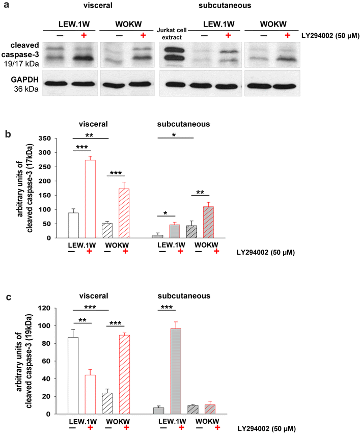
Regulation of the apoptosis marker cleaved caspase-3 under PI3K inhibition. Representative Western blots (a) and corresponding densitometrical analyses of cleaved caspase-3 (b–c). The cleaved caspase-3 expression in adipocyte cell cultures of both strains was slightly detected by Western blot. The inhibition with LY294002 (48 h) caused significantly increase of cleaved caspase-3 expression (17 kDa, mature form of enzyme) in visceral and subcutaneous adipocytes. An extract of Jurkat cells with cytochrome c-induced apoptosis was used in Western blot as the positive control for the occurrence of cleaved caspase-3 (a). Data from n = 6 are presented as mean ± SEM. *p ≤ 0.05, **p ≤ 0.01, ***p ≤ 0.001, according to the one-way analysis of variance together with the Newman–Keuls test. GAPDH was used as normalization control